


Jul
DICOM Explained: How Medical Images Are Stored, Shared, and Managed
Summary
Most people working outside radiology or hospital IT have never heard of DICOM. Honestly, that makes sense it’s not something that comes up in meetings unless something breaks. But if you’ve ever seen a CT scan open on a radiologist’s screen right after the scan finishes, or a report reach a doctor in a different building within minutes, that’s DICOM doing its thing in the background.
The problem it fixes is pretty simple. Hospitals use imaging machines from different companies. Those companies used to build their own formats, their own software, their own way of doing everything. Getting any of it to talk to each other was a nightmare. DICOM came along and said: one format, one standard, everyone uses it.
Introduction
Medical imaging is everywhere in modern healthcare. A patient gets a CT scan in the emergency department, an MRI for a sports injury, or an ultrasound during pregnancy, and those images need to move quickly between scanners, radiologists, clinicians, and hospital systems. The challenge is that these systems often come from different vendors and need to work together without losing critical patient information.
That’s where DICOM (Digital Imaging and Communications in Medicine) comes in. DICOM is the international standard that defines how medical images and related information are stored, transmitted, and managed across healthcare environments. It ensures that a scan produced on one system can be viewed, archived, and shared on another, regardless of the manufacturer.
Today, DICOM sits at the center of almost every imaging workflow. From image acquisition and storage in PACS to teleradiology, cloud imaging platforms, and AI-powered diagnostic tools, it provides the common language that keeps medical imaging systems connected. This article explains what DICOM is, how it works, why it became the industry standard, and how it continues to support modern healthcare infrastructure.
Want to explore the full DICOM standard? The official documentation is available at:
DICOM (Digital Imaging and Communications in Medicine) is the global standard used to store, transmit, manage, and display medical images and associated patient information across healthcare systems.
Objectives
This blog covers the following topics related to the DICOM standard:
- What DICOM is and why it was created
- The structure and contents of a DICOM file
- How DICOM flows through a hospital from registration to report
- The communication services that devices use to talk to each other
- Real-world problems in hospital DICOM deployments
- The modalities and imaging types DICOM supports
- Where DICOM actually shows up in clinical practice
- Where the standard is heading DICOMweb, cloud PACS, FHIR, AI
Overview
So what is DICOM? DICOM stands for Digital Imaging and Communications in Medicine. It’s the global standard for storing, sending, and displaying medical images X-rays, CT, MRI, ultrasound, mammography, and more. But it’s bigger than just the image. A DICOM file carries the patient info, the scan details, the equipment data, and the actual image all in one package.
Before any of this existed, hospitals were mailing physical film. Burning CDs. A scan from a GE machine sometimes wouldn’t open on a Siemens workstation. Radiologists dealt with that constantly. It was slow, it caused errors, and it affected patient care in real ways.
The American College of Radiology and NEMA sat down and built DICOM 3.0, which came out in 1993.
High-Level Component Summary
| Component | Description |
| File Format | Single self-contained package image + patient data + scan metadata |
| Communication Protocol | DIMSE services (C-STORE, C-FIND, C-MOVE, C-GET, C-ECHO) |
| Data Hierarchy | Patient -> Study -> Series -> Instance |
| Web Extension | DICOMweb (STOW-RS, WADO-RS, QIDO-RS) for cloud/browser access |
| Integration Points | PACS, RIS, HIS, EHR, AI systems, teleradiology platforms |
Why It Matters More Than People Think
Nobody appreciates infrastructure until it stops working. DICOM is exactly that kind of thing. A few things it quietly makes possible:
- Any DICOM system can open any DICOM file doesn’t matter who made the scanner or the screen.
- Patient information always travels with the image. You can’t open a scan without knowing who it belongs to.
- Images go from the scanner to the radiologist in seconds, not hours.
- PACS, RIS, HIS they all connect through the same language.
- A study from ten years back can still be pulled up exactly as it was captured.
None of that feels special until you imagine what happens without it.
What’s Inside a DICOM File
A lot of people think it’s basically a JPEG with a few extra details stuck on. It’s really not. The whole file is self-contained image and clinical info are packaged together so nothing ever gets separated.
Inside a typical file: patient name and ID, date of birth, gender, age, the date and time of the scan, the hospital and department, the referring doctor, the modality, and the image itself.
All of that gets stored through DICOM Tags – small identifier codes, each one holding one specific thing:
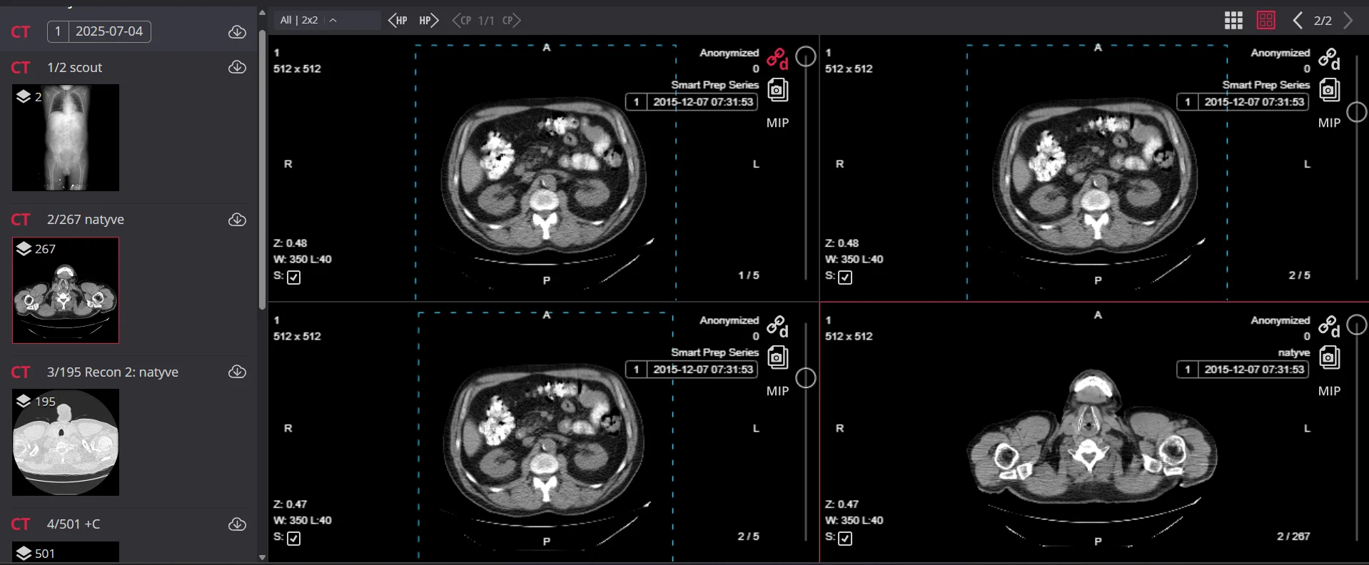
What you see here is a real DICOM viewer displaying multiple CT series. The left panel shows the study hierarchy series by series. Each image on the right carries its own embedded metadata: dimensions, acquisition date, and scan parameters. That’s DICOM working exactly as designed.
For more information visit – DICOMweb
| Tag | What It Stores |
| (0010,0010) | Patient Name |
| (0010,0020) | Patient ID |
| (0008,0020) | Study Date |
| (0008,0060) | Modality |
PACS systems use these tags to sort and find studies on their own. No one is manually logging anything. It just works and when it does, nobody notices, which is kind of the whole point.
The Hierarchy: Patient, Study, Series, Instance
DICOM organises image data in layers. When you’re dealing with hundreds of patients and thousands of images daily, this matters.
| Level | Description | Example |
| Patient | Top-level identifier – one record per person | John Smith, MRN 10042 |
| Study | One full examination | CT Abdomen, Tuesday 14 Jan |
| Series | A distinct phase or protocol within the study | Plain phase, Contrast phase, Delayed phase |
| Instance | One individual image (slice) | Single CT slice -1 of 300 |
Simple structure, but it scales fine whether you’re doing 50 scans a day or thousands.
How It Flows Through a Hospital
Patient comes in. Details go into the RIS or HIS name, ID, exam, referring doctor. Through the DICOM Worklist, that information gets pushed straight to the scanner. The tech sitting down doesn’t type anything again. It’s already there.
Scan happens. DICOM files are created image and patient data together. Those go to the PACS via C-STORE. Archived, indexed, done.
Later the radiologist searches for the study with C-FIND, pulls it with C-MOVE or C-GET. It loads. They report. The referring doctor accesses it from their own desk without ever going to radiology.
That whole chain registration to report runs on DICOM at every single step. Take one piece out and the whole thing breaks.
The Services That Handle Communication
DICOM isn’t just a file format. It’s also a set of services that spell out exactly how devices talk to each other.
| Service | Purpose |
| C-STORE | Moves images from one place to another scanner to PACS, for instance |
| C-FIND | Searches. You give it a name, ID, date range it gives back matching studies |
| C-MOVE | Pulls images and sends them somewhere specific usually a radiologist’s workstation |
| C-GET | Similar to C-MOVE but sends images back to whoever asked, not a third destination |
| C-ECHO | Basic ping IT uses it to check two systems can see each other before anything real happens |
| Modality Worklist | Handles automatic patient data push kills manual entry at the scanner completely |
| Storage Commitment | PACS confirms it actually saved a file safely before the scanner wipes its local copy |
Storage Commitment is the one people miss. Without it, images can disappear during a transfer and you won’t know until someone goes looking.
Common Problems in Real Hospitals
DICOM itself is solid. But real hospital setups are messy old systems, multiple vendors, rushed configs, and sometimes two hospitals merging and bringing all their old problems together.
| Problem | Description |
| Worklist Mismatch | Patient name in the RIS spelled differently than in the modality. Studies end up on the wrong patient or need manual fixes. Even one wrong character is enough. |
| Duplicate Patient IDs | Common after mergers. Two systems combine and one patient exists twice, or two patients share an ID. Pulling a clean imaging history becomes a mess. |
| Transfer Syntax Issues | Not every system handles every compression format. A scan from one modality might not open right on a workstation that doesn’t support that transfer syntax. |
| AE Title Misconfiguration | Devices use short text strings to identify each other. One typo in the setup is enough to kill communication entirely between a modality and the PACS. |
| PACS Migrations | Moving millions of old studies to a new system, without losing metadata or breaking study structure, takes months and still surprises you. |
| Corrupted Studies | Happens when the network cuts out during a transfer. Exactly why Storage Commitment isn’t optional. |
What Modalities It Covers
Basically all of modern imaging:
| Modality Code | Full Name | Primary Use |
| CT | Computed Tomography | Cross-sectional scanning trauma, chest, brain, cancer |
| MRI | Magnetic Resonance Imaging | Soft tissue brain, spine, joints |
| DX / CR | Digital / Computed Radiography | Plain radiography, newer and older digital versions |
| US | Ultrasound | Pregnancy, abdominal, cardiac |
| MG | Mammography | Breast imaging |
| PT | PET | Metabolic activity big in oncology |
| NM | Nuclear Medicine | Functional imaging |
| XA | X-Ray Angiography | Live procedural imaging |
And past those ECG waveforms, endoscopy video, digital pathology. DICOM covers more ground than most people expect.
Where It Actually Shows Up
Teleradiology
A radiologist at home, or in a different country, opens a study from a hospital they’ve never visited. Full quality, no conversion needed. For rural or under-resourced hospitals, this isn’t a bonus feature it’s how they get specialist reads at all.
Emergency Settings
Emergency settings move fast and DICOM keeps up. A trauma scan can be on the emergency physician’s screen within seconds of being done. Nobody’s carrying anything anywhere. It just arrives.
Multi-Site Hospital Networks
A patient scanned at a smaller site has their images ready for review at the main hospital right away no manual file transfers, no waiting.
AI in Radiology
AI in radiology runs on DICOM because that’s what the training data is. Tumour detection, fracture flagging, bleed identification all built to read DICOM directly. One format everywhere means a model built in one place works elsewhere without any conversion step.
Long-Term Archiving
Long-term archiving is a legal thing in most places, and DICOM handles it without drama. Old studies open exactly as they did originally. Nothing degrades or gets lost when systems change.
Where Things Are Heading
| Trend | Description |
| DICOMweb | Built for cloud and browser environments. Runs over HTTP and REST images can be pulled through a regular web browser without special software. What makes cloud PACS and phone-based viewing work. |
| Cloud PACS | Where a lot of new deployments are going. Lower hardware costs, better recovery options, access from anywhere. The data underneath is still DICOM just delivered differently. |
| FHIR Integration | FHIR handles electronic health records. When it connects with DICOM, imaging history and clinical notes end up in the same place. A doctor opens a patient file and sees labs and scans side by side. |
| Structured Reporting with AI | Findings from an algorithm don’t sit in a separate email they get written into the DICOM file itself. Measurements, flagged areas, comparisons to previous studies, all stored with the images permanently. |
| Mobile Access | Mostly sorted now. DICOMweb and modern compression together make it realistic to review diagnostic-quality images on a phone or tablet. On-call from anywhere is already happening. |
Need Help Building Healthcare Imaging Solutions?
Building modern medical imaging platforms requires more than just understanding DICOM. Healthcare organisations need secure, scalable, and interoperable solutions that integrate seamlessly with existing clinical systems.
Ficode helps healthcare providers, medical technology companies, and digital health startups develop:
- DICOM-based imaging solutions
- PACS and vendor-neutral archive (VNA) systems
- Cloud medical imaging platforms
- AWS HealthImaging integrations
- Healthcare interoperability solutions using HL7 and FHIR
- AI-enabled medical imaging workflows
Whether you’re modernising an existing imaging infrastructure or building a new healthcare platform, our healthcare technology specialists can help design and implement solutions that meet clinical, operational, and compliance requirements.
Talk to Our Healthcare Technology Experts
Explore our healthcare solutions – Healthcare
Final Thought
DICOM works best when you don’t notice it. Images load, patient data is attached, a scan from three years back opens fine. That’s the standard doing exactly what it should.
For anyone in PACS, radiology IT, or clinical imaging knowing how it works underneath actually matters. Not because the theory is interesting for its own sake, but because when things go wrong worklist not populating, C-STORE failing, images not reaching the workstation you need to know where to start looking. That always starts with understanding the standard.
Frequently Asked Questions
DICOM (Digital Imaging and Communications in Medicine) is the international standard used to store, transmit, and manage medical imaging data. It ensures that imaging systems from different vendors can exchange information consistently.
DICOM provides a common format and communication standard for medical imaging. Without it, hospitals would struggle to share images between scanners, PACS systems, workstations, and healthcare applications.
DICOM is the standard that defines how medical images and related information are stored and exchanged. PACS (Picture Archiving and Communication System) is the platform that stores, manages, and displays those DICOM images.
DICOM combines a file format with a communication protocol. It stores images together with patient and study information and enables systems to exchange that data using services such as C-STORE, C-FIND, C-MOVE, and C-GET.
DICOM supports a wide range of imaging modalities, including CT, MRI, X-ray, ultrasound, mammography, PET, nuclear medicine, angiography, and many other healthcare imaging formats.
DICOMweb is a modern web-based extension of DICOM that uses REST APIs and HTTP protocols. It enables cloud imaging platforms, browser-based image viewers, mobile access, and integration with modern healthcare applications.
Yes. Modern cloud imaging solutions use DICOM and DICOMweb to store, retrieve, and share medical images securely across healthcare organizations and remote locations.
Most medical imaging AI systems use DICOM data for training and analysis. Standardized image formats and metadata make it easier to develop, deploy, and integrate AI solutions into clinical workflows.

Nihal Rajput is the Operations Director at Ficode Software Solutions Pvt. Ltd., where he oversees day-to-day operations, streamlines processes, and drives organisational efficiency. With a focus on delivering scalable solutions and maintaining operational excellence, he plays a key role in aligning teams, optimising resources, and ensuring client satisfaction as the company grows.

 Previous
 Next
Next